





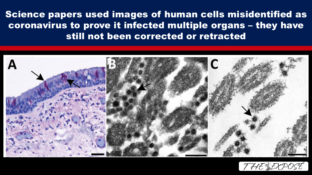
A 2021 review by CDC scientists found that 23 out of 27 electron microscopy reports from March–November 2020 misidentified common human cellular structures as coronavirus.
These misidentified particles were published in top medical journals as evidence of direct SARS-CoV-2 infection in organs like the liver, heart, kidney and intestine.
The structures were mistaken for virus because their size and protein fringes mimicked the characteristic viral spikes.
The CDC researchers warned that these errors could propagate, as “articles with misidentified viral particles are used by others to verify the presence of viral particles in their own research.”
Several of these papers remain published without retractions or corrections withdrawing the disputed electron-microscope identifications.
Let’s not lose touch…Your Government and Big Tech are actively trying to censor the information reported by The Exposé to serve their own needs. Subscribe to our emails now to make sure you receive the latest uncensored news in your inbox…
Top Medical Journals Label Images of Human Cell Structures as ‘Coronavirus’—‘Misidentified Viral Particles Are Used by Others to Verify the Presence of Viral Particles’: CDC
By Jon Fleetwood, 8 September 2026
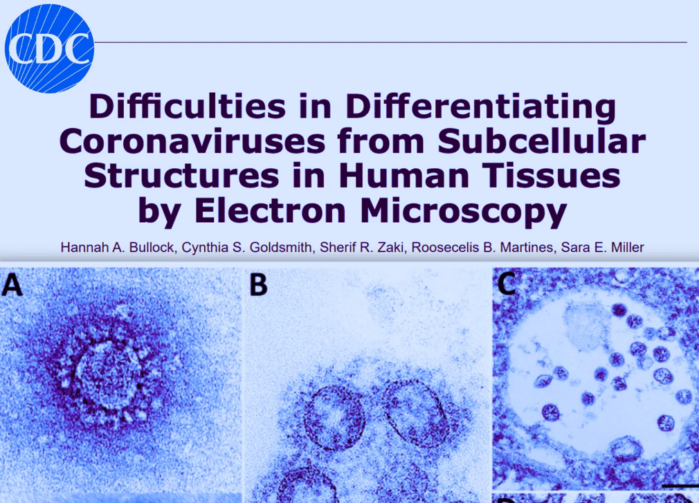
Top medical journals published images of ordinary human cellular structures labelled as “coronavirus,” according to a 2021 review authored by Centres for Disease Control and Prevention (“CDC”) scientists and collaborators titled ‘Difficulties in Differentiating Coronaviruses from Subcellular Structures in Human Tissues by Electron Microscopy’.
The researchers examined 27 reports that used electron microscopy to identify coronavirus directly in human tissue and found that 23 contained cellular structures misidentified as virus.
“In each case of erroneously identified coronavirus particles,” they wrote, “the structures mistaken for virus are common cellular organelles.”
More than five years later, several of the papers remain published without corrections withdrawing the disputed electron-microscope identifications, including studies claiming direct SARS-CoV-2 infection of the kidney, liver, heart, intestine and other organs.
The development raises questions about how much of the historical record describing the purported virus’s physical presence and morphology rests on images that were never uniquely identifying.
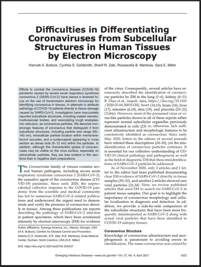
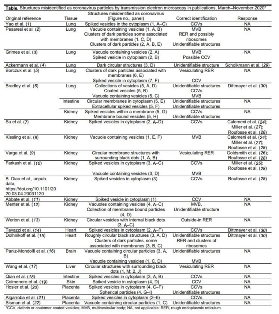
Human Cell Structures Published as “Coronavirus”
The CDC-associated researchers published their review in Emerging Infectious Diseases in April 2021.
They had searched reports published between March and November 2020 that used electron microscopy to identify coronavirus directly in patient specimens.
They found 27 reports with electron microscopy findings. “The other 23 articles revealed a pattern of subcellular structures misidentified as virus,” they wrote.
Those structures included clathrin- and coatomer-coated vesicles, multivesicular bodies, rough endoplasmic reticulum, and other nonviral structures.
Some had been used to support much larger claims about covid-19.
A 2020 Journal of the American Society of Nephrology paper, for example, was titled ‘Ultrastructural Evidence for Direct Renal Infection with SARS-CoV-2’.
The University of Michigan researchers said electron microscopy revealed particles “morphologically identical to SARS-CoV-2” and concluded that they provided evidence of “productive direct infection of the kidney.”
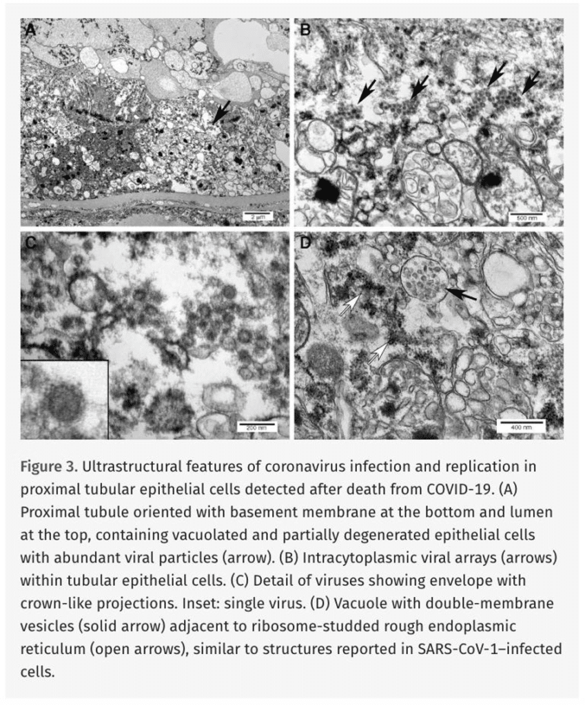
The CDC review later classified the pictured “spiked vesicles” as coated vesicles and the purported virus-filled vacuole as a multivesicular body.
The paper remains published.
Its formal correction changed reported particle measurements from micrometres to nanometres.
It did not withdraw the disputed identification of the structures as SARS-CoV-2.
“Coronavirus” in Liver and Heart Studies Was Human Cell Structures
The same problem reached the liver.
A 2020 Journal of Hepatology study titled ‘SARS-CoV-2 infection of the liver directly contributes to hepatic impairment in patients with COVID-19’ reported finding what its authors called “typical coronavirus particles” in liver cells.
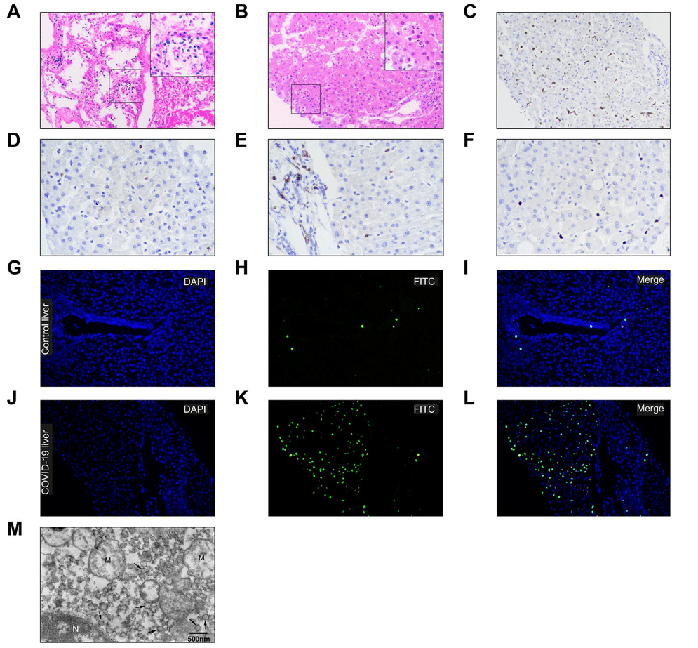
The electron-microscope images were part of the evidence used to argue that SARS-CoV-2 directly infected the liver.
The CDC-associated review subsequently identified the pictured circular structures as vesiculating rough endoplasmic reticulum, not coronavirus.
The original paper remains in the scientific record under its direct-infection title.
Similar findings appeared in the heart.
A paper in the European Journal of Heart Failure titled ‘Myocardial localisation of coronavirus in COVID-19 cardiogenic shock’ reported that biopsy examination “demonstrated” viral particles in heart tissue.
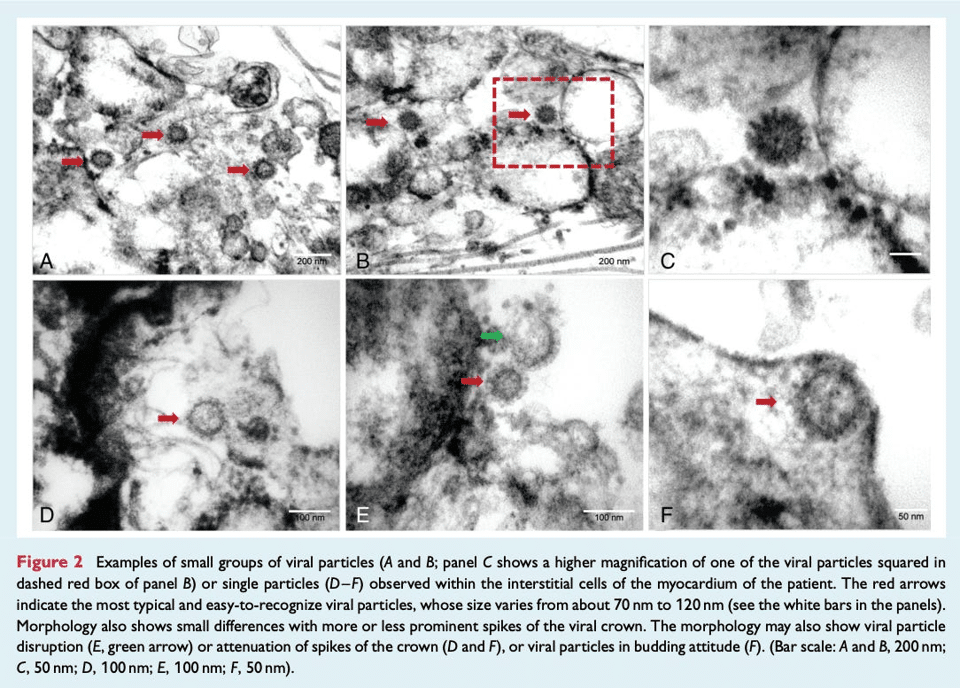
The CDC review subsequently classified the pictured spiked vesicles as clathrin-coated vesicles.
That paper also remains published.
“SARS-CoV-2 Virions” in Intestine Study Were Cellular Vesicles
In July 2020, Clinical Infectious Diseases published an article titled ‘Direct Evidence of Active SARS-CoV-2 Replication in the Intestine’.
“Typical SARS-CoV-2 virions in the intestinal epithelial cells under electron microscopy were observed,” the authors wrote, saying the images “provided direct evidence” of active replication.
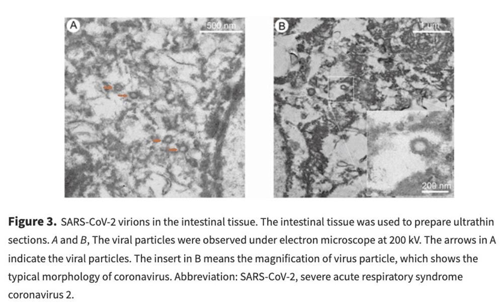
The CDC review later classified the pictured “spiked vesicles” as clathrin-coated vesicles.
The journal page remains online under the “Direct Evidence” title.
Human Cell Structures Can Meet “Morphological Criteria for a Coronavirus”
The CDC review explained how human cellular structures could acquire the appearance attributed to coronavirus.
Clathrin-coated vesicles are circular cellular structures measuring approximately 60–100 nanometres. They are surrounded by a fringe of clathrin protein.
“The clathrin or coatomer projections protruding from the vesicles as a fringe can be easily misinterpreted as viral spikes,” the researchers wrote.
Multivesicular bodies posed another identification problem because both they and what researchers classify as coronavirus accumulations can appear as membrane-bound collections of spherical particles.
Rough endoplasmic reticulum could also form circular structures with attached ribosomes resembling purported spikes.
The overlap could be remarkably close.
The CDC-associated researchers described another cellular structure approximately 100 nanometres in diameter that they said “meet[s] the morphological criteria for a coronavirus” except that its internal dots measured approximately 20 nanometres instead of the 6–12 nanometres they interpreted as cross-sections of viral nucleocapsid.
“The exact nature of their composition or relationship to any cellular processes has not been determined,” they wrote.
CDC Warned the Errors Could Rewrite the Record
The researchers warned that once incorrectly identified images entered the literature, they could become references used to validate later purported virus images.
“Articles with misidentified viral particles are used by others to verify the presence of viral particles in their own research,” they wrote. That could result in researchers “wrongfully documenting the presence of the virus in damaged tissues.”
The fuller context of those quotes is revealing.
The CDC researchers write in their conclusion:
The issue of virus misidentification within tissue samples is not unique to coronaviruses or limited to those subcellular structures we addressed in this report; nuclear pores may be mistaken for herpesvirus, perichromatin granules for smaller DNA viruses such as parvoviruses or polyomaviruses, and ribosomes for picornaviruses. Neurosecretory granules and glycocalyceal bodies can also be misidentified as viruses.
In each case of erroneously identified coronavirus particles, the structures mistaken for virus are common cellular organelles. These misinterpretations are easy to make without extensive training and are made easier by the publication of incorrectly identified viral structures. Articles with misidentified viral particles are used by others to verify the presence of viral particles in their own research, potentially wrongfully documenting the presence of the virus in damaged tissues. However, rather than actual virus infection of a failing organ, the damage could be due to lack of support of the infected organ due to the body’s response to toxins, such as cytokines and circulating debris, or to clotting. These continued misinterpretations could have meaningful impacts on future publications about coronavirus detection and research.
Bullock HA, Goldsmith CS, Zaki SR, Martines RB, Miller SE. Difficulties in Differentiating Coronaviruses from Subcellular Structures in Human Tissues by Electron Microscopy. Emerg Infect Dis. 2021;27(4):1023-1031. https://doi.org/10.3201/eid2704.204337
The warning wasn’t hypothetical.
By the time the CDC review was published, disputed images had already appeared in papers claiming direct infection of multiple human organs.
And years later, several of those papers remain part of the scientific record without corrections withdrawing the electron-microscope evidence the CDC-associated researchers challenged.
That raises a larger question about the historical record of SARS-CoV-2 itself: How much of the early scientific literature describing where the purported virus was physically present in the human body (and what researchers said it looked like) rests on images that were never uniquely identifying?
Bottom Line
CDC scientists and collaborators found that human cellular structures had been published as “coronavirus” across 23 reports, including papers claiming direct SARS-CoV-2 infection of major human organs.
The structures included ordinary cellular components whose size, shape and protein fringes could satisfy characteristics researchers attributed to coronavirus, including structures the CDC-associated researchers said could “meet the morphological criteria for a coronavirus.”
The researchers warned that published misidentifications could be used by other scientists to validate their own images, “potentially wrongfully documenting the presence of the virus in damaged tissues.”
More than five years later, several of the papers containing those disputed images remain in the scientific record without corrections withdrawing the electron-microscope identifications.
That leaves an unresolved problem at the foundation of the historical SARS-CoV-2 record: human cellular structures identified by CDC-associated electron microscopists remain published as evidence of where the purported virus was physically present in the human body, while the CDC researchers themselves warned that such misidentifications could propagate through subsequent research.
More from Jon Fleetwood:
- ‘Majority’ of covid Genomes Had ‘Systematic Errors’ as Computers Turned Missing Sequence Data Into False Evidence of SARS-CoV-2 Evolution: Journal ‘Nature Methods’
- SARS-CoV-2’s Origin Story Relies on a Coronavirus Reference Library Built by the Same Gov’t-Funded Network That Planned SARS-CoV-2-Like Viruses Before the Pandemic
- The Self-Referencing Closed Loop of Coronavirus Genetics Began With Assumptions About Marks From Radioactive Material Scientists Themselves Added
- US Government Has Funded Ralph Baric’s Coronavirus Work and Patents Since 1984
- Baric Predecessor and ‘Father of Coronavirus Research’ Michael Lai Assigned Inferred Genetic Traits to Coronavirus Without Observing Them Directly
- NIH Hands Up to $2 Billion in Future Pandemic, Other Emergency Projects to Pentagon Funding Vehicles GAO Still Cannot Track
- BioNTech Kills mRNA Cancer Shot Phase 2 Study After Finding ‘More Deaths in the Vaccine Arm’ – 10 Months After Futility Warning
- Pfizer-Type Lipid Nanoparticles Even Containing No mRNA Increase Metastatic Tumour Burden More Than 10-Fold in Mice With Cancer: Journal ‘Nano Today’
- FDA Approves Pfizer’s New XFG covid Shot Without Human Safety or Efficacy Trial of New Formula—Genetic Sequence Hidden From Public, Raising Informed Consent Alarms
- FDA Approves Moderna XFG covid Vaccines 81 Days Before Teen/Adult Safety Study, 126 Days Before Children’s Study
- The Self-Referencing Closed Loop of Coronavirus Genetics Began With Assumptions About Marks From Radioactive Material Scientists Themselves Added
- WHO Says Pandemic Determinations Can Be Made Without ‘Evidence of Illness’
- Former Senior NIAID Official Pleads Guilty to ‘Conspiracy to Commit Offenses and to Defraud the United States’ Regarding covid-19 Origins
- PHEMCE Centralised 7 Federal Agencies and Private Industry Into ‘Holistic, End-to-End’ Pandemic-Industrial Complex
- HHS Funds Chimeric Bird Flu Pandemic Vaccine Made with Engineered Plague Derivative BECC470s: Journal ‘mSphere’
- Trump/Kennedy FDA Approves Moderna mRNA-1010 Influenza Shot ‘mFLUSIVA’ with Less Than 1% Absolute Benefit and 6 Times More Severe Reactions
- 2018 DARPA/EcoHealth ‘DEFUSE’ Proposal Documented All Three SARS-CoV-2 Spike Anomalies Before the Pandemic
- DARPA’s Pandemic Stack: Building the Pandemic Operating System Before covid
- Beyond Lab Leak and Natural Spillover: The ‘In Silico Theory’ of Pandemic Orchestration
About the Author
Jon Fleetwood is an American investigative journalist, author and independent analyst known for his work on health policy, biotechnology and political narratives. He publishes articles on his Substack page ‘Jon Fleetwood’. You can also follow him on Instagram HERE and Twitter (now X) HERE.
He is the author of ‘An American Revival: Why American Christianity Is Failing & How to Fix It’ and co-author of ‘What We’re Afraid to Ask: 365 Days of Healing for Adult Survivors of Childhood Abuse’.
The Expose Urgently Needs Your Help…
Can you please help to keep the lights on with The Expose’s honest, reliable, powerful and truthful journalism?
Your Government & Big Tech organisations
try to silence & shut down The Expose.
So we need your help to ensure
we can continue to bring you the
facts the mainstream refuses to.
The government does not fund us
to publish lies and propaganda on their
behalf like the Mainstream Media.
Instead, we rely solely on your support. So
please support us in our efforts to bring
you honest, reliable, investigative journalism
today. It’s secure, quick and easy.
Please choose your preferred method below to show your support.

Categories: Breaking News, Latest News, World News
I still think that Muslims have infiltrated the CDC and the FDA in the USA because they are exempt from getting covid shots.
Interesting. Didn’t know that. How many do?
AI says Islam approved of the shots (halal) and muslims could only be excused on an individual basis, like everybody else.
Anyone whose sincere religious belief does not allow then to accept a vaccine or other medical product has an exemption under Title 7 of the Civil Rights Act of 1964.
The person does not have to be a formal member of any organized religious group, but the belief must be sincere and be a principle or rule that guides how they live their life.
“(j) The term “religion” includes all aspects of religious observance and practice, as well as belief, unless an employer demonstrates that he is unable to reasonably accommodate to an employee’s or prospective
employee’s religious observance or practice without undue hardship on the conduct of the employer’s business.”
“SEC. 2000e-2. [Section 703]
(a) Employer practices
It shall be an unlawful employment practice for an employer –
https://www.eeoc.gov/statutes/title-vii-civil-rights-act-1964
For example, some object to receiving a product ino their body which was developed using and/or contains aborted human fetal cells or fragments- which the products labeled as “COVID-19 Vaccine” all do.
Others object to the commercialization of parts of aborted babies, such as cell lines used for research, development, and manufacture of medical products, including the “COVID-19 Vaccine”. The us of such products makes the killing of developing babies a for-profit business.
More information on our Rights regarding the products which were covered by the PREP Act:
https://CovidPenalty.com
‘Knowledge is Power.’
Here in the USA all of congress exempted themselves but not their constituents also, What did they know?
That was illegal because the Constitution requires all of us to be treated the same when law is applied, not I must do x but you don’t.
MDs were exempt when nurses and everybody else working in medical care were not. Many who refused lost their jobs while the docs working elbow to elbow with them did not.
I hate articles like this. You make the fact that there are fake Viral photos…true enough. But then you talk about “Viral Signatures” that are real. This implies there are things such as a Covid19 VIRUS…. No there is not. There is not ONE actual verified photo of a “Viral Pathogen” None… EVER.
While I agree with you that the whole ‘field’ of “Virology” appears to be a massive fakery and fraud – where they use the same kinds of techniques as exposed in this article to fabricate or fantacise the existence of invisible, unproven viruses, one question remains.
What is the true cause of so many so-called viral illnesses? E.g. Influenza, the common cold… These illnesses do appear to be contagious, but if it’s not viruses, bacteria or other infectious organisms, what is the mechanism? Why do people get so sick, especially around changes of the seasons?
And please don’t give me “Terrain Theory”, unless you can justify it properly. I have never been able to have a successful discussion with a Terrain Theorist that’s any better than “virology”. I ask them specific questions and they can’t answer.
Read Dr. Marizelle Arce’s brilliantly researched and extremely well presented book ‘Germs Are Not Our Enemy.’ (2025).
And there are your answers.
Her work is also on Substack.
GermsAreNotOurEnemy.com
Exactly right. The pictures they use as proof of a virus, is dead cell debris. Cells die and break up all the time and when there is a lot of dead cells breaking down at the same time, the body has to detoxify which can make you feel yuk and give you symptoms they call “flu”. Dr Samantha Bailey explains this well in her youtube videos as well in her book “Viru Mania”, which I can highly recommend you read.
No surprise here. The bad guys will use every possible tool to lie to us. Electron microscopy is one of the useful tools.
Does anyone remember the post-doc researcher at Cambridge who, in 2020 or 2021, surreptitiously obtained a vial of covid virus and tested its contents? He did a youtube video about it. He was sorely puzzled because he totally believed scientific officialdom’s narrative but could not find any covid virus whatsoever in the vial, no matter what test he used. I wonder where he is now, sweeping floors in some basement, if they let him live?
More likely to have been in 2019. My memory for dates is perfect.
I meant imperfect. My typing isn’t perfect either.
Not one single virus has ever been proven to exist. The pictures they claim to be viruses are mere dead cell debris. It’s a scam that has been allowed to carry on for decades, so big pharma can profit from vaccines.